Gout CT Scan: How DECT Can See Uric Acid Crystals That Blood Tests and X-rays Miss
Recurring joint pain, sudden swelling in the big toe, or repeated gout flare-ups can be difficult to explain when routine tests appear inconclusive. Many patients continue experiencing symptoms despite “normal” uric acid levels or unremarkable X-rays. This is one of the key reasons gout is often underdiagnosed or detected late.
Advanced imaging is changing that. A specialised gout CT scan using Dual Energy CT technology can directly identify uric acid crystal deposits that traditional tests may fail to detect. By offering greater precision and earlier detection, DECT is helping clinicians diagnose gout more confidently and guide treatment more effectively.
In many cases, DECT imaging helps reveal:
- Hidden uric acid crystal deposits before joint damage becomes visible
- Crystal accumulation within tendons and soft tissues missed on routine imaging
- The difference between gout and other inflammatory joint conditions
- Uric acid kidney stones that require a different treatment approach
Why Traditional Gout Tests Have Limitations
Gout develops when excess uric acid forms crystals inside joints and surrounding tissues, triggering inflammation and severe pain. While blood tests and X-rays remain important, they do not always provide definitive answers.
Blood tests measure uric acid levels, but these readings can fluctuate. Some patients experiencing active gout attacks may still show normal levels during testing. X-rays, meanwhile, mainly detect structural damage that appears later in the disease process.
This diagnostic gap often delays timely treatment.
| Diagnostic Method | What It Detects | Limitation |
| Blood Test | Uric acid levels | May appear normal during flare-ups |
| X-ray | Bone and joint damage | Early gout may not be visible |
| Ultrasound | Inflammation and deposits | Operator-dependent findings |
| DECT gout imaging | Uric acid crystal deposits | Highly detailed and precise |
A gout diagnosis scan using DECT offers a more advanced solution because it identifies the crystal deposits themselves rather than relying only on indirect signs.
What Is a Gout CT Scan?
A gout CT scan uses specialised Dual Energy CT technology to detect uric acid crystal deposits inside joints, tendons, and soft tissues.
Unlike conventional CT imaging, DECT captures images using two different energy levels. This allows radiologists to distinguish uric acid from calcium and bone structures with high accuracy. The result is colour-coded imaging that clearly highlights crystal accumulation.
In clinical practice, gout CT imaging is especially valuable when symptoms persist, but routine investigations remain inconclusive.
What DECT Can Detect
A dual energy CT scan for gout can identify:
- Early uric acid crystal deposits
- Tophi or visible gout nodules
- Crystal build-up in tendons and soft tissues
- Multiple affected joints during one scan
This detailed evaluation helps clinicians assess both disease activity and long-term crystal burden more accurately.
Why DECT Is Becoming Important in Gout Diagnosis
For many years, joint fluid aspiration was considered the standard method for confirming gout. Although effective, it involves inserting a needle into the affected joint to collect fluid for microscopic analysis.
This is where DECT for gout offers a major advantage. It is non-invasive, fast, and capable of directly visualising uric acid deposits.
Key Benefits of DECT Imaging
Earlier diagnosis
A uric acid CT scan can reveal deposits before permanent joint damage develops, allowing earlier intervention.
Better diagnostic clarity
Symptoms of gout may resemble rheumatoid arthritis, osteoarthritis, or infection. DECT improves differentiation between these conditions.
Monitoring treatment response
Imaging can help assess whether uric acid deposits are reducing after medication and lifestyle changes.
Evaluation of complex cases
Patients with persistent pain, recurrent flare-ups, or unclear blood test results often benefit from advanced imaging.
Can a CT scan Detect Gout Reliably?
A common question patients ask is: can a CT scan detect gout accurately?
A routine CT scan alone is not designed specifically for gout detection. However, a specialised gout CT scan radiology assessment using DECT technology can identify and map uric acid crystal deposits with high precision.
Doctors may recommend DECT imaging when:
- Blood tests remain inconclusive
- Joint aspiration is not feasible
- Symptoms continue despite treatment
- Multiple joints are involved
- Hidden crystal deposits are suspected
Because DECT visualises the crystals directly, it offers a higher level of diagnostic confidence in difficult cases.
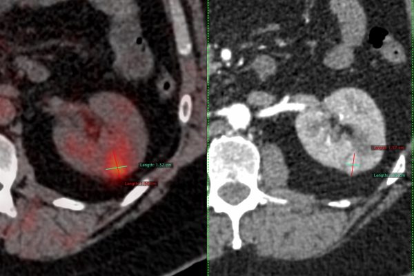
Can DECT Detect Uric Acid Kidney Stones?
Patients frequently ask: do uric acid stones show on CT?
Yes. Dual Energy CT can identify uric acid kidney stones and distinguish them from calcium stones. This distinction is important because uric acid stones may sometimes dissolve with medication, while calcium stones often require different treatment strategies.
Similarly, many patients search: do uric acid kidney stones show up on CT scan if standard imaging is unclear. DECT improves stone characterisation and supports more precise management decisions.
Who Should Consider a Gout Diagnosis Scan?
Not every patient with joint pain requires advanced imaging. However, a gout imaging test may be recommended when symptoms persist despite routine investigations.
You may benefit from DECT imaging if you experience:
- Recurrent gout attacks
- Persistent swelling or stiffness
- Inconsistent uric acid test results
- Suspected tophi or hidden deposits
- Kidney stones linked to elevated uric acid levels
Early detection matters because untreated gout can eventually lead to chronic inflammation and irreversible joint damage.
Conclusion
Gout diagnosis is not always straightforward. Blood tests and X-rays may fail to identify crystal deposits during the early stages, leaving patients with recurring symptoms but limited clarity.
A specialised gout CT scan using DECT technology offers a more advanced and accurate approach by directly visualising uric acid crystal deposits within joints and tissues. From early diagnosis to treatment monitoring and kidney stone evaluation, DECT is becoming an increasingly valuable tool in modern gout care.
If ongoing joint pain, swelling, or unexplained flare-ups continue to affect daily life, advanced imaging may provide the answers routine tests have missed. Connect with Picture This by Jankharia to explore whether DECT imaging could support a faster and more precise diagnosis.
Frequently Asked Questions
A specialised dual energy CT scan for gout can identify uric acid crystal deposits directly, making it highly effective in confirming gout when symptoms or blood tests are unclear.
- It detects crystal deposits before visible joint damage develops
- It improves diagnostic accuracy in recurrent or atypical cases
Yes. DECT gout imaging is more sensitive than standard X-rays because it detects uric acid deposits rather than only structural joint damage.
- It helps distinguish gout from other inflammatory joint disorders
- It identifies disease earlier in the diagnostic process
A uric acid CT scan highlights crystal deposits within joints, tendons, and surrounding soft tissues using colour-coded imaging.
- It can detect hidden crystal accumulation not visible on routine scans
- It supports better treatment planning and disease monitoring
Yes. Patients may experience active gout despite normal uric acid levels during testing, which is why a gout diagnosis scan can be valuable.
- Blood uric acid levels can fluctuate during flare-ups
- Imaging helps confirm whether crystal deposits are present
Yes. DECT imaging can identify uric acid kidney stones and distinguish them from calcium stones, helping guide the right treatment strategy.
- Some uric acid stones may dissolve with medication
- Accurate stone identification helps prevent recurrence