Role of Whole-Body PSMA PET in Detecting Rare Metastatic Sites: Bone, Liver, and Lymph Nodes
Prostate cancer has a well-known tendency to spread to the bones and lymph nodes, but it can occasionally extend to less common sites such as the liver or distant soft tissues. Identifying these metastases early is critical, as it directly influences staging, prognosis, and treatment decisions.
Traditional imaging methods, such as bone scans, CT scans, and MRI, though useful, often miss small or atypically located lesions, especially in the early stages. These techniques primarily detect structural changes, which may not appear until the disease has progressed.
Whole-body PSMA PET imaging, including PSMA PET/CT, represents a significant advancement in this area. By combining molecular-level sensitivity with full-body coverage, it allows for the detection of even subtle metastatic activity, including rare or unexpected sites. This comprehensive approach not only improves diagnostic accuracy but also ensures that patients receive more precise and personalised care.
Why “Whole-Body” Matters
Most PSMA PET studies focus on the pelvis and torso, aiming to identify the typical areas of spread. However, metastases can sometimes extend beyond the standard field into distal bones, the lower extremities, or soft tissues. A whole-body PSMA PET scan extends the field of view from head to toe, increasing the chance of detecting disease.
- In one series using total-body ^68Ga‑PSMA‑11 PET/CT, about 8.5% of patients had bone metastases outside the usual scanning range. These extra-range lesions led to changes in staging or treatment in many cases.
- Another study found that by extending acquisition beyond mid-thigh, additional bone lesions were identified in ~6% of patients (though in that cohort, it rarely changed management).
- Additionally, rare metastases to soft tissues (e.g., liver, lung, adrenal glands, brain) have been documented more readily with PSMA PET than with conventional imaging.
Thus, whole-body coverage reduces “blind spots” and can unmask rare metastatic sites.
Detecting Bone Metastases: How PSMA PET Shines
The bone is the most common site for metastatic prostate cancer. But not all bone lesions are equal: some are subtle, in the marrow or located far from usual scanning windows.
Advantages of PSMA PWT in bone detection
- PSMA PET is more cell-specific than bone scans, which detect bone remodelling (which could be because of trauma, arthritis, infection, etc.). That means fewer false positives from non-cancer bone activity.
- In comparative studies, ^18F‑PSMA tracers outperform conventional bone scans for sensitivity and specificity in detecting skeletal metastases.
- Whole-body extension captures lesions in distal sites (e.g., foot). A case report describes a patient whose 18F‑PSMA PET/CT (scanned only to mid-thigh) missed foot metastases; they were only revealed by a post-therapy ^177Lu-PSMA scan covering the full body.
Cautions to keep in mind
- PSMA uptake is not 100% specific to prostate cancer: benign bone processes (fractures, degenerative changes, Paget’s disease) may show uptake. Clinical context and correlating CT/MRI features help.
- Very small bone lesions might still escape detection if tracer uptake is faint or resolution is limited.
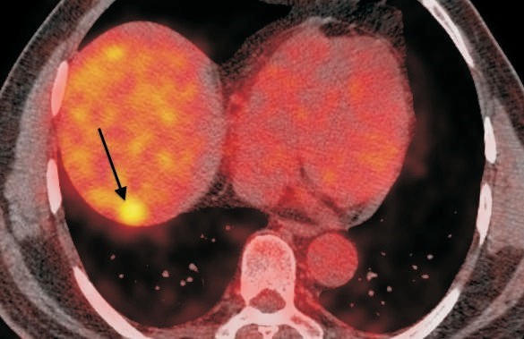
Liver Metastases: Rare But Important
Metastasis to the liver from prostate cancer is relatively uncommon (reported in ~4–5% of cases) but carries significant prognostic weight.
How whole-body PSMA PET helps
- PSMA PET can detect small liver lesions that might be asymptomatic or missed on CT/MRI. In rare case series, liver metastases have been identified by Ga‑68 PSMA PET/CT in otherwise unsuspected patients.
- By covering the entire torso, including the upper abdomen, whole-body imaging ensures that metastases near the diaphragm or upper lobes are not missed.
Challenges in liver detection
- The liver naturally shows physiological uptake of PSMA tracers (moderate baseline activity), which can mask or complicate the interpretation of subtle lesions.
- Lesions with low PSMA expression may remain invisible. Some prostate cancer clones downregulate PSMA, requiring complementary imaging modalities (e.g., FDG PET, MRI).
Lymph Nodes: From Pelvis to Distant Fields
Lymphatic spread is the second most frequent route of metastasis in prostate cancer, after bone. Standard imaging often focuses on pelvic, iliac, and retroperitoneal nodes. However, PSMA PET’s whole-body scope can detect distant nodal disease (such as supraclavicular, mediastinal, and inguinal).
Strengths in lymph node detection
- PSMA PET is markedly superior to conventional CT/MRI in detecting small, sub-centimetre metastatic lymph nodes—even those without overt enlargement. One recent study showed PSMA PET/CT improved detection of nodal metastases from 13.3% (CT alone) to 56.7%.
- Whole-body imaging captures unexpected nodal spread—cases of inguinal nodal metastasis, solitary inguinal or inguinal-to-abdominal wall, or even supraclavicular nodes have been documented.
- Because PSMA PET is molecular, even nodes with minimal enlargement but harbouring microscopic disease can light up.
Interpretation cautions
- Non-prostate causes of PSMA uptake (e.g., benign nodes, inflammation) need caution; clinical correlation is essential.
- Very small PSMA-negative metastatic nodes may still slip away from detection.
Comparison of Popular Imaging Modalities in Cancer Diagnosis
| Imaging Modality | Type of Imaging | Strengths | Limitations | Common Uses |
| CT scan | Cross-sectional X-ray | Quick, widely available, good for anatomy | Radiation exposure, limited soft tissue contrast | Detecting tumours, staging, checking organ spread |
| MRI | Magnetic resonance | Excellent soft tissue detail, no radiation | Expensive, slower, not suitable for metal implants | Brain, spinal cord, pelvic, liver tumours |
| PET Scan | Molecular imaging | Shows metabolic activity, good for staging | Limited anatomical detail, expensive | Detecting active tumours, assessing recurrence |
| Ultrasound | Sound waves | Safe, real-time, portable | Operator-dependent, limited depth/clarity | Guiding biopsies, examining superficial organs |
| X-ray | Basic radiography | Fast, cheap, good for bones and lungs | Poor soft tissue contrast | Bone lesions, chest abnormalities |
Clinical Impact & Decision-Making
The revelation of rare or unsuspected metastases can change a patient’s staging from localised to systemic, prompting different therapeutic strategies (e.g., systemic therapy, inclusion in trials, radiation field extension, etc.). Studies have shown that PSMA PET/CT findings changed treatment plans in about 28–45% of patients in some cohorts.
Key considerations for clinicians:
- When to do a whole-body PSMA PET scan? In high-risk disease, biochemical recurrence, or when conventional imaging is equivocal.
- Complementary modalities (FDG PET, MRI, DWI, or CT) may catch PSMA-negative metastases. A study combining whole-body DWI with PSMA PET/MR found additional lesions (mostly lymph nodes) in ~36% of patients.
- Histopathologic confirmation may be needed for unusual metastatic sites if the result would alter management (e.g., a liver lesion that might be another primary).
- Scanning protocol: ensure that imaging covers from skull vertex to toes when clinically justified, to avoid missing extra-range disease.
Frequently Asked Questions
It’s a PSMA PET/CT (or PET/MR) imaging approach that covers the entire body—from head to toes—so that metastases outside the usual pelvis-to-thigh field are not missed.
Unlike CT, MRI, or bone scans, which reflect anatomy or bone remodelling, PSMA PET uses a radiotracer that binds prostate cancer cells (or cells expressing PSMA). This molecular imaging is more specific and sensitive for detecting small metastases.
Yes. F‑18–labelled PSMA tracers typically have a longer half-life, higher image resolution, and more favourable logistics (wider distribution) compared to Ga‑68 tracers. Many clinical centres now favour F‑18 PSMA agents for these reasons.
No. Some metastases may have very low PSMA expression, be below the detection limit, or be masked by physiologic uptake. That’s why combining PSMA PET with MRI, DWI, or other modalities is often useful.
Not always—if the imaging findings are convincing and consistent with the disease pattern, treatment may proceed. However, if confirming the nature of the lesion would change management (e.g., distinguishing a second primary cancer or benign lesion), a biopsy may be warranted.
It tends to be more resource-intensive because of extended acquisition time, tracer usage, and technical requirements. But the “cost” is balanced against the value of more precise staging and better-informed therapy decisions. Over time, technology advances, and wider adoption may narrow that gap.
Whole-body PSMA PET scans have reshaped how we detect and manage prostate cancer, especially when it spreads beyond common areas. Unlike conventional imaging, this advanced technique scans from head to toe, revealing rare metastatic sites in the bones, liver, and lymph nodes that might otherwise go unnoticed. This broader view leads to more accurate staging and better-informed treatment decisions.
At Picture This, by Jankharia, we’re committed to offering cutting-edge diagnostic tools, such as the F-18 PSMA PET scan, to ensure that no detail is missed. By combining precision with full-body coverage, we help doctors see the complete Picture—so patients get the care they truly need when they need it most.

