Cryoablation of Pelvic Fibromatosis
Cryoablation of Pelvic Fibromatosis
Fibromatosis is a difficult tumor to treat in many instances. If superficial and small, it can be surgically removed. However, if the lesion is large, infiltrative or in deep areas, surgery can be morbid and may still be inadequate.
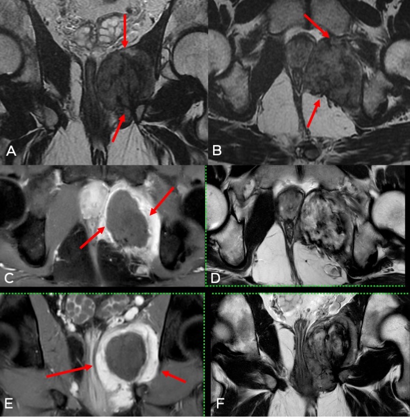
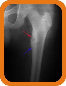
In such instances, alternate methods such as certain forms of chemotherapy and ablation techniques such as radiofrequency ablation (RFA) and cryoablation have been successfully used (Fig. 1).
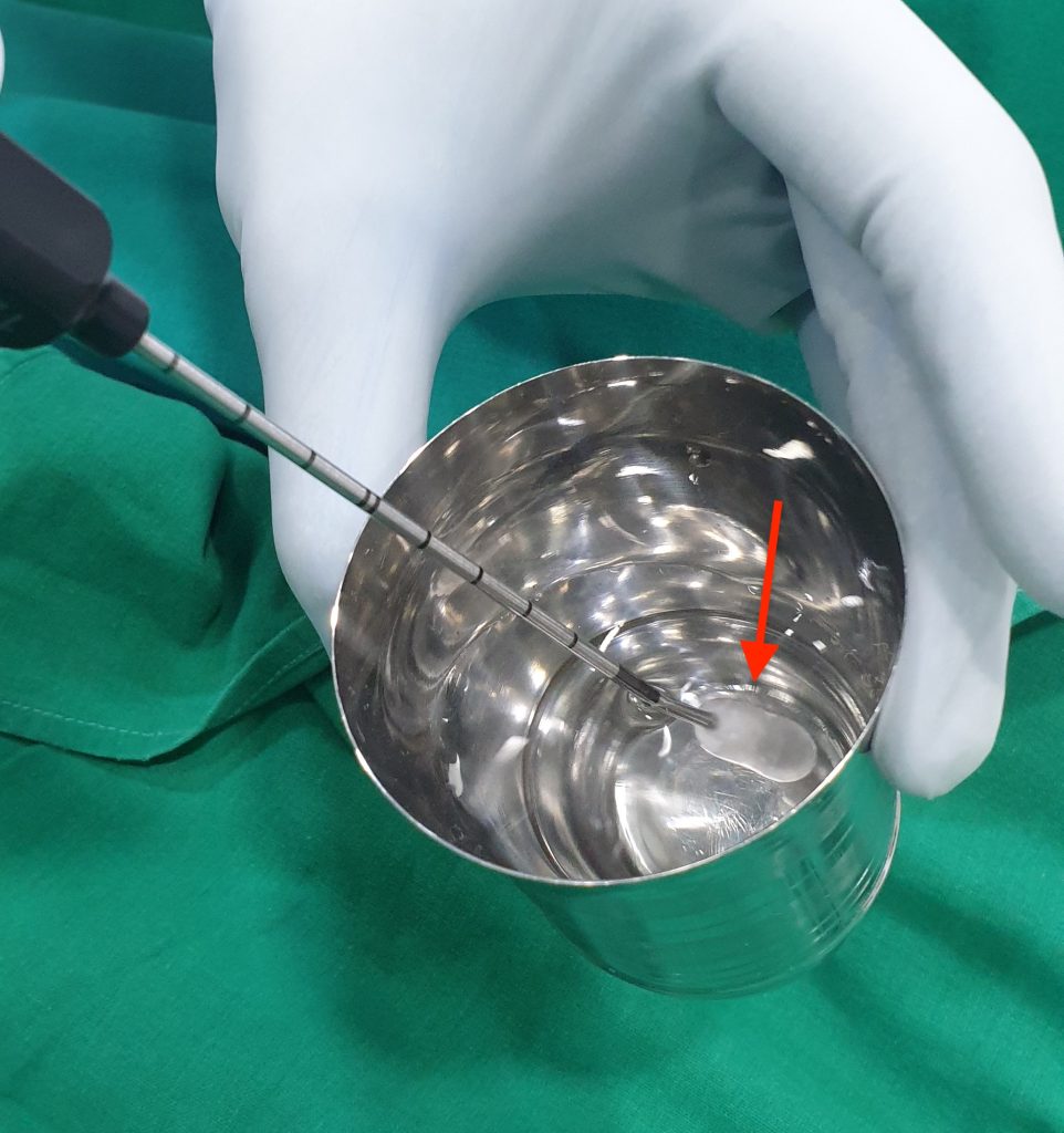
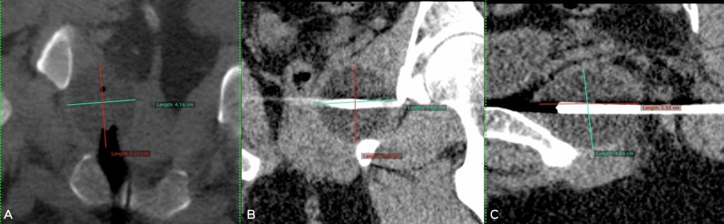
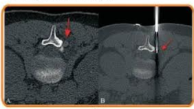
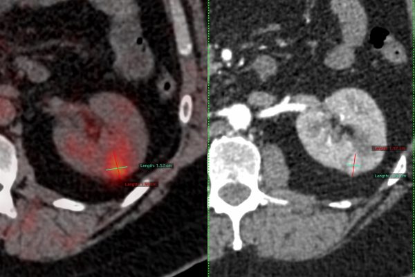
Cryoablation has been shown to be the better ablation technique compared to others. Cryoablation is a relatively new technique where freezing temperatures are used to kill tumor cells by creating iceballs within the tumors. Liquid nitrogen or argon is used depending on the system. With liquid nitrogen, the temperature at the centre of the ice-ball can reach up to minus 196 degrees Celsius with lethal temperatures of minus 20 to minus 40 degrees in the rest of the iceball (Fig. 2). More importantly, the iceball can be visualized on USG, CT or MRI as the case may be, allowing accurate monitoring of the ablation area (Fig. 3).
The machine is compact and sits in the CT scan room. The procedure is easily done on an out-patient basis in day-care, mostly under local anesthesia; some patients need intravenous sedation if the tumors are inherently painful.
Indications
- Bone tumors
- Soft tissue tumors like fibromatosis (Fig. 1) for cure and palliation
- Lung tumors – primary and metastases
- Liver tumors – primary and metastases.
- Renal tumors
Contradiction
- Large tumor size (typically > 10 cm)
- Abnormal coagulation profile