Cryoablation of Chondroblastoma
Cryoablation of Chondroblastoma
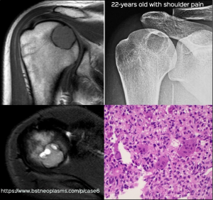
Chondroblastoma is a benign bone tumor that contains chondroblastic cells. It typically occurs in the age group of 10-25 years and 75% occur in the epiphysis of the long bones (Fig. 1).
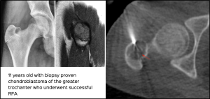
Chondroblastomas were treated in the past with curettage. Over the last 10 years, radiofrequency ablation (RFA) has become a popular method for treating chondroblastomas less than 3.0 cm in size (Fig. 2).
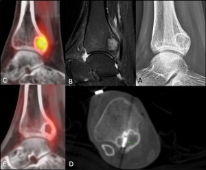
Cryoablation is expected to have similar results [1] and causes less post-procedure pain than RFA and is an alternative to RFA when it comes to benign bone tumors like chondroblastoma (Fig. 3), osteoid osteomas and osteoblastomas.
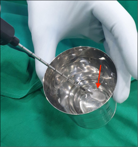
Cryoablation treatment is a relatively new technique where freezing temperatures are used to kill tumor cells by creating iceballs within the tumors. Liquid nitrogen or argon is used depending on the system. With liquid nitrogen, the temperate at the centre of the ice-ball can reach up to minus 196 degrees Celsius with lethal temperatures of minus 20 to minus 40 degrees in the rest of the iceball (Fig. 4).
 Indications
 Indications
- Bone tumors – chondroblastomas, osteoid osteomas, osteoblastomas
- Soft tissue tumors like fibromatosis (Fig. 1) for cure and palliation
- Lung tumors – primary and metastases
- Liver tumors – primary and metastases.
- Renal tumors
Contraindications
- Large tumor size
- Abnormal coagulation profile
Points:
- Cryoablation is a new ablation technique that uses ice-balls to freeze and kill tumor cells
- Chondroblastoma < 3.0 cm in size are often treated with RFA.
- Cryoablation is a good alternative to RFA
Legends
Fig. 1: Humeral chondroblastoma. Typical X-ray, MRI and histopathology findings of a humeral chondroblastoma.
Fig. 2: RFA of chondroblastoma. 11-years old with a biopsy proven chondroblastoma underwent successful radiofrequency ablation (RFA).
Fig. 3 (A-E). Cryoablation of chondroblastoma. X-ray (A), sagittal T2 MRI (B) and PET (C) show typical features of a distal tibial chondroblastoma in a 20-years old. A 3-3-5 freeze-thaw-freeze cycle was used during the cryoablation (D) with a 13 G probe and intravenous sedation. The post-procedure PET (E) done 10 days later shows complete lack of FDG uptake. The patient was asymptomatic.
Fig. 4: In-vivo iceball during the procedure.