MRI for Sports Injuries: Diagnosing ACL Tears, Meniscus Damage & Rotator Cuff Injuries
You twist your knee during a football match, feel a sharp pain while changing direction, and suddenly find it difficult to continue playing. Or perhaps shoulder pain after a gym session refuse to settle despite rest. Sports injuries can affect anyone, from professional athletes to weekend fitness enthusiasts. The challenge is knowing exactly what has been injured and how serious it is.
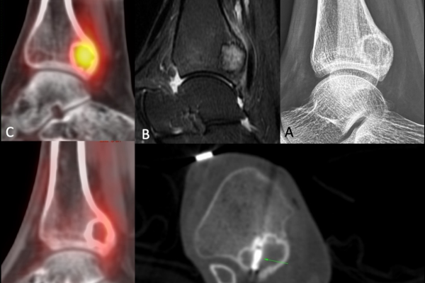
This is where MRI for sports injuries becomes valuable. MRI provides detailed images of ligaments, tendons, cartilage, muscles, and joints, helping doctors identify injuries accurately and plan the right treatment.
Why MRI Is Important for Sports Injury Diagnosis
Many sports injuries involve soft tissues rather than bones. While X-rays are useful for identifying fractures, they cannot clearly show ligaments, tendons, cartilage, or muscles.
An athletic injury imaging assessment using MRI helps doctors evaluate these structures in detail. MRI uses magnetic fields and radio waves to create high-resolution images without radiation exposure.
This level of detail is particularly useful when symptoms such as pain, swelling, instability, or weakness do not clearly point to a single injury.
What MRI Can Detect
- Ligament injuries: Tears and sprains affecting joint stability.
- Tendon damage: Partial or complete tendon tears.
- Cartilage injuries: Damage that may affect movement and cushioning.
- Muscle injuries: Strains, tears, and inflammation.
- Bone bruises: Hidden injuries not visible on standard X-rays.
ACL Tears and the Role of MRI
The anterior cruciate ligament, commonly called the ACL, is one of the main stabilising ligaments in the knee. ACL injuries often occur during football, basketball, tennis, and other sports involving sudden twisting or pivoting movements.
Typical symptoms include swelling, pain, and a feeling that the knee may give way.
How MRI Helps Diagnose ACL Tears
MRI confirms whether the ligament is partially or completely torn. It also identifies associated injuries that may affect treatment decisions.
| MRI Finding | What It Indicates | Clinical Significance |
| Partial ACL tear | Some fibres remain intact | May respond to rehabilitation |
| Complete ACL tear | Full ligament disruption | Surgery may be considered |
| Bone bruise | Impact injury within the knee | Suggests significant trauma |
| Meniscus injury | Additional cartilage damage | May require separate treatment |
Patients often search for ACL tear diagnosis MRI cost when considering imaging. While cost is a factor, the value of MRI lies in providing the detailed information needed to guide recovery and treatment planning.
Meniscus Tears: Why Accurate Imaging Matters
Within the knee, the menisci help provide cushioning and stability by reducing friction between the bones during movement. Meniscus tears commonly occur during twisting movements or high-impact sports activities.
Symptoms can include pain, swelling, clicking sensations, and difficulty fully straightening the knee.
What MRI Shows in Meniscus Injuries
Meniscus tear imaging allows radiologists to determine the location, size, and pattern of the tear. This information is important because treatment options depend on the type of injury.
| Tear Type | MRI Appearance | Possible Management |
| Small stable tear | Limited cartilage disruption | Physiotherapy and monitoring |
| Bucket handle tear | Displaced cartilage fragment | Often requires surgery |
| Degenerative tear | Wear-related changes | Individualised treatment |
| Complex tear | Multiple tear patterns | Specialist evaluation |
MRI can also reveal whether other knee structures have been injured at the same time, which is common in sports-related trauma.
Rotator Cuff Injuries and Shoulder MRI
Shoulder injuries are frequently seen in swimmers, cricketers, tennis players, and weightlifters. The rotator cuff consists of a set of muscles and tendons that provide shoulder stability while supporting a wide range of arm movements.
When these tendons become injured, everyday activities such as lifting, reaching, or throwing may become painful.
When Is a Rotator Cuff MRI Needed?
Doctors may recommend a rotator cuff tear MRI when shoulder pain persists despite treatment, weakness develops, or a significant injury has occurred.
What MRI Reveals
- Tendon inflammation: Early tissue changes before major damage occurs.
- Partial tears: Small areas of tendon injury.
- Full-thickness tears: Complete disruption of the tendon.
- Muscle changes: Long-standing injuries affecting muscle quality.
This detailed assessment helps doctors decide whether physiotherapy, medication, injections, or surgery may be the most appropriate option.
When Should You Consider an MRI?
Not every sports injury requires advanced imaging immediately. Minor strains and sprains often improve with rest and rehabilitation.
However, MRI may be recommended if you experience:
- Persistent pain despite treatment
- Significant swelling
- Joint instability
- Reduced range of motion
- Difficulty bearing weight
- Suspected ligament or tendon injury
- Recurrent injuries in the same area
Early imaging can often shorten the time taken to reach a clear diagnosis.
What to Expect During the Scan
MRI is a non-invasive and painless procedure.
During the examination:
- You will lie on a scanning table.
- The injured body part will be positioned carefully.
- The scanner produces tapping sounds during image acquisition.
- Remaining still helps produce clear images.
- Most musculoskeletal MRI scans take between 20 and 45 minutes.
After the scan, an experienced radiologist reviews the images and prepares a detailed report for your referring doctor.
How MRI Supports Better Treatment Decisions
MRI helps doctors understand not only whether an injury exists but also how severe it is. This information supports more accurate treatment planning and can help avoid unnecessary procedures.
For patients seeking MRI for sports injuries Mumbai, high-quality imaging provides valuable clarity when symptoms are limiting daily activities, work, or sporting performance.
Take the Next Step Towards a Clear Diagnosis
Ignoring persistent pain after a sports injury can delay recovery and increase the risk of further damage. If symptoms continue despite rest or treatment, an MRI can provide the detailed information needed to understand the problem. At Picture This by Jankharia, advanced MRI technology helps deliver accurate diagnosis and timely clinical guidance. Early answers can help you move forward with confidence.
Don’t leave your recovery to guesswork.
Frequently Asked Questions
MRI is one of the most reliable imaging methods for assessing ligaments, tendons, cartilage, and muscles. As part of an athletic injury imaging assessment, it provides detailed information that helps doctors identify the extent of an injury and plan treatment effectively.
Yes. MRI is highly effective for confirming knee cartilage injuries. MRI for sports injuries can show the location and severity of a meniscus tear, helping determine the most appropriate management approach.
Not always. However, when significant ligament damage is suspected, meniscus tear imaging and MRI evaluation can help identify associated injuries and support accurate treatment planning.
A rotator cuff tear MRI can reveal inflammation, partial tears, complete tears, and muscle changes around the shoulder. These findings help doctors decide on the most suitable treatment pathway.
Patients looking for MRI for sports injuries Mumbai should choose a centre with advanced imaging equipment and experienced musculoskeletal radiologists. Accurate imaging plays a key role in achieving a precise diagnosis and recovery plan.