Seeing Spinal Instability Clearly
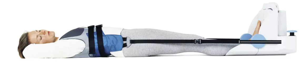
At Picture This, we offer advanced Stress MRI of the spine, leveraging our Philips 3T Ingenia CX system paired with specialized stress devices. This dynamic imaging captures the spine under load and relaxation-essential for diagnosing conditions that static scans might miss.
What Is Stress MRI of the Spine?
A Stress MRI (sometimes called weight-bearing or kinematic MRI) involves imaging the lumbar spine in different positions-such as flexion and extension-to highlight mechanical instability, facet joint cysts, spondylolysis, or Modic changes. Unlike standard MRIs, stress imaging reproduces the spine's behaviour under movement, improving detection of dynamic conditions.
Why Choose Picture This for Spine Stress MRI?
- Philips 3T MRI with Stress Device - High-resolution, dynamic imaging.
- Expert Radiologists - Including Dr. Bhavin Jankharia, Dr Vaishali Nimbkar, Dr Shilpa Sankhe, Dr Karthik Ganesan, Dr Nidhi Doshi
- Comprehensive Imaging Suite - MRA, elastography, MARS MRI, CT for cross-validation.
- Comfort First - Spacious bore, ergonomic positioning, attentive monitoring.
- Fast Turnaround - Reports delivered by the next day, or sooner for urgent cases.
- Collaborative Interpretation - We liaise directly with your spine specialist or surgeon.
When Is Stress MRI Recommended?
Stress MRI is invaluable when you have:
- Unexplained low back pain worsened by movement.
- Suspected facet cysts or instability causing intermittent issues.
- Pars interarticularis stress reaction or early spondylolysis, common in athletes
- Modic changes-signal alterations in vertebral endplates linked to mechanical stress and pain.
- Cases where standard supine MRI fails to capture dynamic pathology.