3D Mammography (Digital Breast Tomosynthesis) in Mumbai – Advanced Breast Imaging at Picture This
Seeing Breast Tissue in Unprecedented Detail
At Picture This by Jankharia, we believe early detection can change everything. That’s why we’ve invested in 3D Digital Breast Tomosynthesis, the most advanced mammography technology available today. It gives us sharper, clearer images of breast tissue, helping us find even the smallest abnormalities earlier and with greater confidence.

What is 3D Mammography?
3D mammography, also known as digital breast tomosynthesis, is cutting-edge imaging technology that provides a more detailed view of breast tissue.
Instead of a single flat image like traditional 2D mammography, this technique takes multiple low-dose X-ray pictures from different angles. A computer then reconstructs these into thin, layered slices similar to turning the pages of a book.
By reducing the effect of overlapping tissue, 3D mammography allows radiologists to detect smaller tumors, subtle changes, or hidden abnormalities with greater accuracy and confidence.
Why Choose 3D Mammography at Picture This?
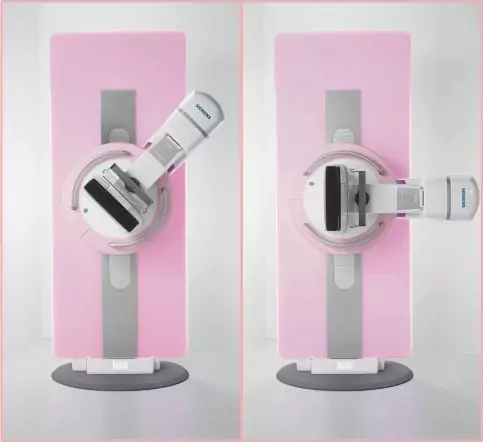
1. State of the Art Siemens Inspiration 3D Unit
One of the first full-field digital tomosynthesis machines in India, equipped with stereotactic biopsy capability.
2. Subspecialized Breast Radiologist
Dr. Bijal Jankharia leads our breast imaging team, bringing national recognition for expertise in mammography and ultrasound correlation.
3. Superior Detection in Dense Breasts
3D mammography reduce false negatives and recall rates—critical for women with dense glandular tissue.
4. Comfort-Focused Design
Padded compression paddles and optimized positioning minimize discomfort without compromising image quality.
5. Access to high-resolution ultrasound in the same visit for comprehensive evaluation.
Clinical Benefits of Digital Breast Tomosynthesis
1. Higher Cancer Detection Rates
Studies show up to 30% more invasive cancers detected compared to 2D alone.
2. Fewer Callbacks
Layer-by-layer review decreases false positive findings and reduces patient anxiety.
3. Improved Characterization
Better visualization of lesion margins and 3D architecture.
4. Guided Biopsy and Vaccum Assisted Biopsy (VAB)
Stereotactic biopsy attachments enable precise sampling of tiny or deep lesions.
Who Should Consider 3D Mammography?
While every woman can benefit, we especially recommend it for:
- Women over 40 for annual breast screening.
- Women with dense breast tissue.
- Women with a personal or family history of breast cancer.
- Women who have had inconclusive results from previous mammograms.
- Women with breast symptoms like lump, pain, nipple discharge etc…