The Concept of Benign Aggressive Bone Tumors

Download PDF

The Concept of Benign Aggressive Bone Tumors

Bone tumors are best evaluated with a combination of plain radiographs and MRIs. There are various signs and rules that differentiate benign from malignant lesions and help us arrive at a specific diagnosis.

One such is the concept of the “benign aggressive” lesion. Clinically, these are defined as benign tumors with locally aggressive behavior. Radiologically, these are defined as tumors that appear benign on X-rays but aggressive on MRI with marrow and perosseous edema, effusion and periosteal reaction, but without a soft tissue mass.

The following conditions present as “benign” on X-rays and “aggressive” on MRI. They can be further differentiated based on age, appearance and location

  1. Osteoid osteoma / Osteoblastoma (Fig. 1)
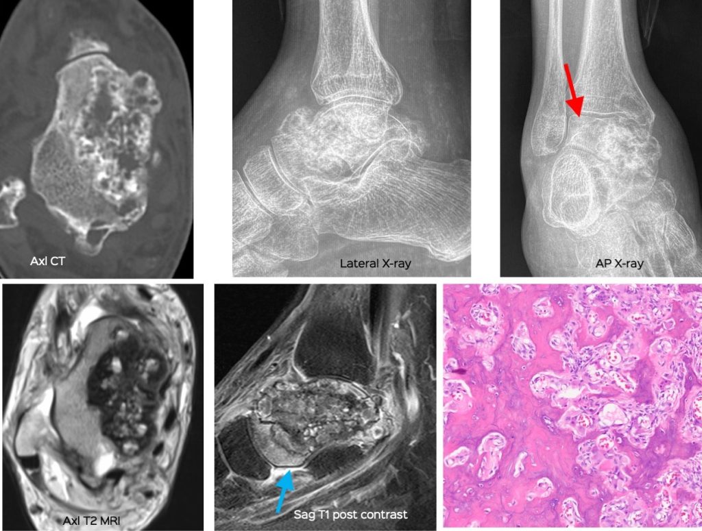
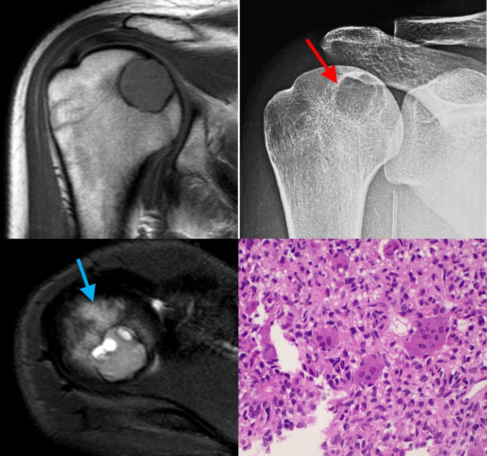
  2. Chondroblastoma (Fig. 2) / Chondromyxoid fibroma
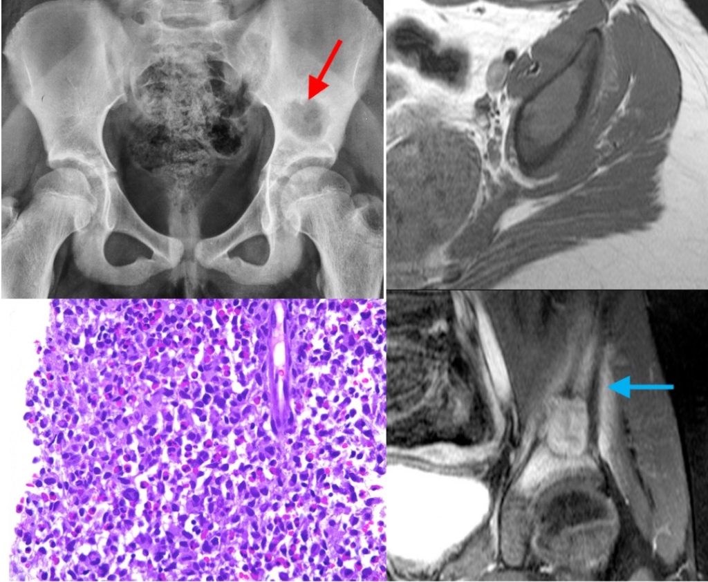
  3. Langerhans cell histiocytosis (Fig. 3)
  4. Benign tumors with fractures
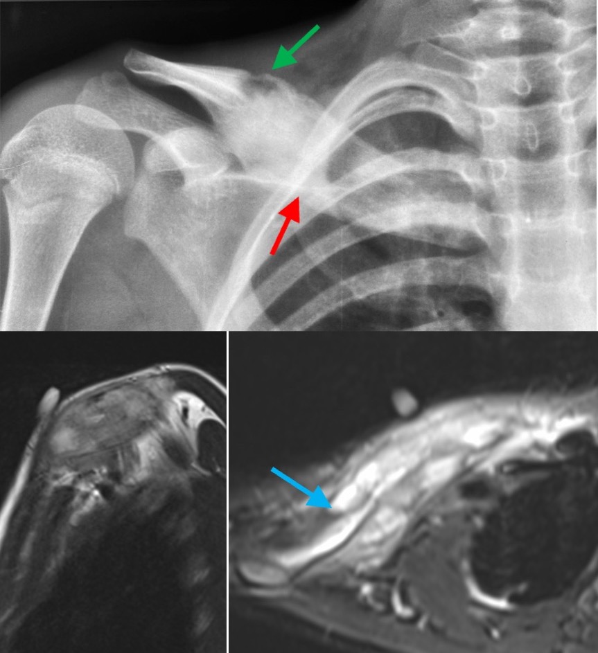
  5. Chronic nonbacterial osteitis (Fig. 4)
  6. Giant cell tumor / Aneurysmal bone cyst

These tumors should be differentiated from infection / osteomyelitis, which can simulate “benign aggressive” lesions superficially.

Points:

  • Bone tumors or focal bone lesions can be difficult to diagnose and characterize
  • Many signs and rules help
  • Lesions that are “benign” on X-rays and “aggressive” on MRI are few and narrow the differential diagnosis.
the-concept-of-benign-aggressive-bone-tumors-3
Fig. 1: Osteoblastoma. 60-years old man shows a bone-forming lesion in the talus. On the X-rays, it has a narrow zone of transition with a sclerotic rim (red arrow). On the MRI, there is marrow edema (blue arrow), but no soft tissue mass. This was correctly diagnosed to be osteoblastoma, confirmed on histology.
the-concept-of-benign-aggressive-bone-tumors-4
Fig. 2: Chondroblastoma. 22-years old has a focal osteolytic lesion in the epiphysis of the proximal humerus, showing a sclerotic rim (red arrow) on the X-ray. The MRI shows marrow edema (blue arrow), which is sign of aggression. This location and appearance are typical of chondroblastoma, which was confirmed on histology.
the-concept-of-benign-aggressive-bone-tumors-5
Fig. 3: Langerhans cell histiocytosis. 11-years old has a focal osteolytic lesion with a narrow zone of transition (red arrow) in the left iliac bone. The MRI shows marked marrow edema (blue arrow). The biopsy showed Langerhans cell histiocytosis (LCH).
the-concept-of-benign-aggressive-bone-tumors-6
Fig. 4: Chronic nonbacterial osteitis. 12-years old has a right clavicular pathology with diffuse cortical thickening and periosteal reaction (red arrow) and a small focal osteolytic lesion (green arrow). The MRI shows an aggressive lesion with marrow edema (blue arrow). This is typical of chronic nonbacterial osteitis – two biopsies were negative for a definite etiology.