The Role of Imaging Techniques in Guiding Cryoablation for Fibromatosis
Introduction:
Cryoablation has emerged as a promising non-invasive treatment option for fibromatosis, offering effective tumor control with minimal invasiveness. One key aspect that enhances the success of cryoablation is the utilization of advanced imaging techniques. These imaging modalities play a crucial role in guiding the procedure, ensuring accurate tumor targeting, and monitoring the treatment process. In this blog post, we will explore the significance of imaging techniques in guiding cryoablation for fibromatosis, shedding light on their contribution to successful outcomes and improved patient care.
Understanding Fibromatosis and Cryoablation:
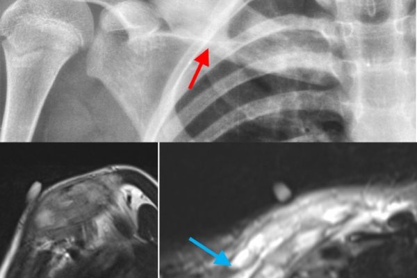
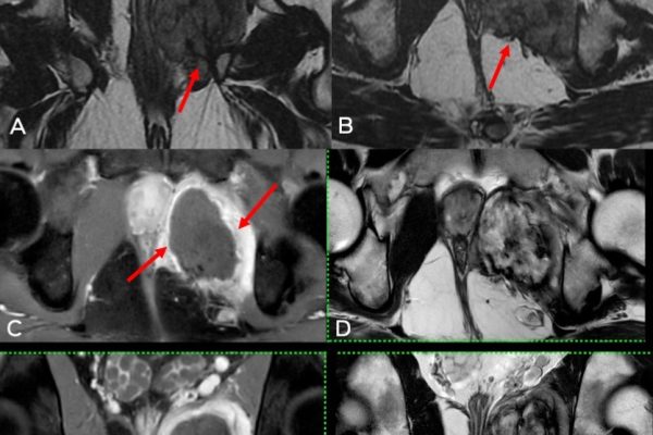
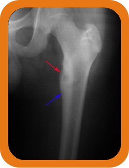
Fibromatosis, also known as desmoid tumors, is a rare condition characterized by the growth of benign but aggressive tumors in the connective tissues. Cryoablation is a minimally invasive treatment approach that utilizes extremely cold temperatures to destroy these abnormal tissues. During cryoablation, precise tumor targeting is critical to ensure effective tumor destruction while minimizing damage to surrounding healthy tissues. This is where imaging techniques come into play, offering real-time visualization and guidance throughout the procedure.
Ultrasound-Guided Cryoablation:
Ultrasound imaging is commonly used to guide cryoablation procedures for fibromatosis. It provides real-time imaging of the tumor and surrounding structures, allowing physicians to accurately position the cryoprobe and monitor the freezing process. Ultrasound can visualize the size, location, and boundaries of the tumor, enabling precise targeting. It also helps assess the extent of tumor destruction during the procedure. The ability to visualize ice formation and monitor its growth allows physicians to adjust the cryoablation parameters, ensuring optimal treatment outcomes.
CT Scan-Guided Cryoablation:
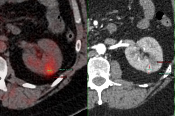
In addition to ultrasound, computed tomography (CT) scans can also be utilized to guide cryoablation procedures for fibromatosis. CT scans provide detailed cross-sectional images of the tumor and surrounding structures, offering precise anatomical localization. CT scans can assist in accurate needle placement and monitoring the positioning of the cryoprobe during the procedure. The use of CT scans allows for improved visualization of the tumor and helps assess treatment response. By combining CT scan guidance with ultrasound, physicians can enhance accuracy in tumor targeting and treatment monitoring.
Combining Imaging Techniques:
In some cases, a combination of imaging techniques may be utilized to guide cryoablation procedures for fibromatosis. For instance, ultrasound can be used initially for real-time guidance during needle placement, followed by a CT scan to provide more detailed imaging and evaluation of the tumor. This multimodal approach allows for enhanced accuracy and confidence in tumor targeting and treatment monitoring. By leveraging the strengths of each imaging modality, physicians can maximize the benefits of cryoablation while minimizing potential risks.
Advantages and Limitations:
The utilization of imaging techniques in guiding cryoablation for fibromatosis offers several advantages. It enhances procedural accuracy, improves tumor targeting, and helps in real-time monitoring of the treatment process. Imaging guidance also allows for minimal invasiveness, reducing the risk of complications and promoting quicker recovery. However, it is important to note that each imaging modality has its own limitations, such as cost, availability, and patient factors. The choice of imaging technique depends on various factors, including tumor characteristics, equipment availability, and physician expertise.
Conclusion :
Imaging techniques play a vital role in guiding cryoablation for fibromatosis, enabling accurate tumor targeting and treatment monitoring. Ultrasound and CT scans provide real-time visualization and enhance procedural accuracy, leading to improved outcomes and patient care. As technology advances and imaging capabilities improve, the role of imaging techniques in cryoablation will continue to evolve, contributing to further advancements in fibromatosis treatment.